THE NEUROVASCULAR UNIT IN STROKE RECOVERY
Most stroke survivors suffer moderate to severe long-term functional deficits despite the treatment availability of tissue plasminogen activator and endovascular thrombectomy. Potentiating the beneficial effects of rehabilitation therapy in the chronic stage of stroke thus remains a major clinical challenge. We study the acute neurovascular sequelae following focal cortical ischemia to design better tools to promote long term recovery.
To this end, we use multi-scale and multi-modal imaging (MRI, fUS, 2PLSM and PET) and electrophysiology (laminal and multi electrode arrays) to study the long term benefits of treatments on neurovascular recovery following stroke. By imaging neuronal and vascular collapse in real time before, during, and after ischemia we can examine the efficacy of different therapeutic approaches on neurovascular coupling and use Machine Learning to build models that predict the trajectory of the recovery.
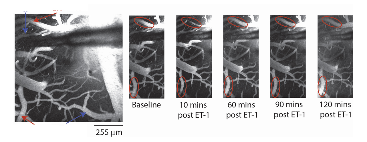
Two-photon fluorescence microscopy of cortical vasculature surrounding the site of vasoconstrictor injection showing temporal propagation of vessel collapse in stroke. Red arrows indicate arteries, and blue arrows indicate veins.
Activity map for stroke murine neurovasculature. The colour gradient depicts relative activity percentages for individual vessels over the 20 second acquisition period.