tools
MULTIPHOTON LASER SCANNING MICROSCOPE SYSTEMS

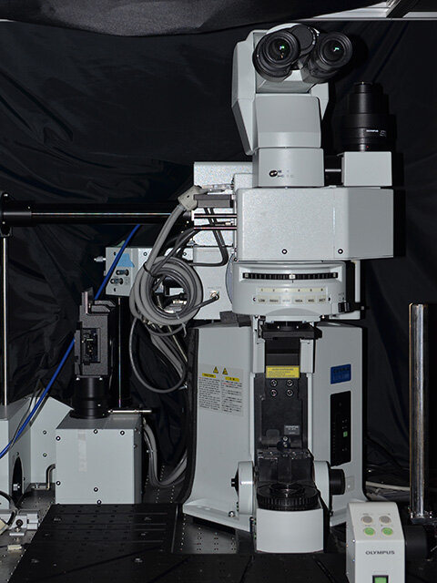
Olympus FVMPE-RS
The newer FVMPE-RS system allows high speed scanning (up to 30 frames per second for a 512x512 full field of view; or 438 frps at 512x32) with an optical path optimized for near-infrared transmission, allowing deeper imaging into the brain. Light is sourced from two Spectra Physics’ Titanium:sapphire lasers (MaiTai and Insight models for concurrent deep red imaging (680-1300nm) and photostimulation for optogenetic applications. The microscope is also equipped with visible light photostimulation capabilities (458 and 552nm), sequential scan management for complex imaging paradigms, and triggering systems for integration with other lab equipment. Find more technical information here.
Olympus FV1000MPE
The Olympus FV1000MPE upright multiphoton laser scanning microscopy system is equipped with an IR Mai Tai HP mode-locked Titanium Sapphire DeepSee tunable laser (690–1040 nm, Spectra Physics) for multiphoton imaging and with visible laser lines (405 nm, 440nm, 488nm, 514nm, 561n, and 635nm) for confocal imaging. Emitted flourescence is collected using four external photomultiplier tubes (Hamamatsu Photonics). The system is used to scan fixed or live images, and features include Z-stack 3D imaging and time series up to 16 frames/sec. Find full technical information here.
PRECLINICAL MAGNETIC RESONANCE IMAGING AND SPECTROSCOPY SYSTEMS

Bruker BioSpec 70/30 USR with Avance III HD
Ultra-shielded refrigerated horizontal bore 7T scanner
Gradient system capable of 200mT/m of gradient amplitude with inserts for rats and mice
Avance III HD digital radio frequency system capable of multi-channel transmitting and receiving (up to 16)
Integrated shimming up to second order
Range of RF coils suited for a wide range of applications - volume coil, phased-array receive-only surface coil, planar receive-only surface loop coil and more
Find more supplier technical information here

Bruker Ascend 300WB with Avance III HD
Ultra-shielded vertical wide bore 7T magnet optimized for NMR
Water-cooled gradient system capable of 1.5T/m @ 60A with imaging coils covering sample sizes of 5 - 25 mm
Integrated shimming up to fourth order
Range of RF coils suited for a wide range of applications – 1H, 13C, and 31P volume coils with quadrature transmission, planar receive-only
Find more supplier technical information here
Both systems are equipped with ParaVision software (PV 6.0.1) that comes with application packages/pulse sequences and tools for analysis and visualization of fMRI, perfusion, metabolism, diffusion, and spectroscopy.
ELECTROPHYSIOLOGY SETUP

A-M Systems Model 3600 16-Channel Extracellular Amplifier
Two sixteen channels amplifiers (AM3600) with stimulating and recording capabilities and 32-channel data acquisition card to use in combination with Multi Electrode Arrays (MEAs) and Linear Microelectrode Arrays (LMAs) for Local Field Potentials (LFPs) and Multi Unit Activity (MUAs) recordings. Find complete technical information here.

PRECLINICAL HIGH FREQUENCY MICRO ULTRASOUND IMAGING SYSTEM
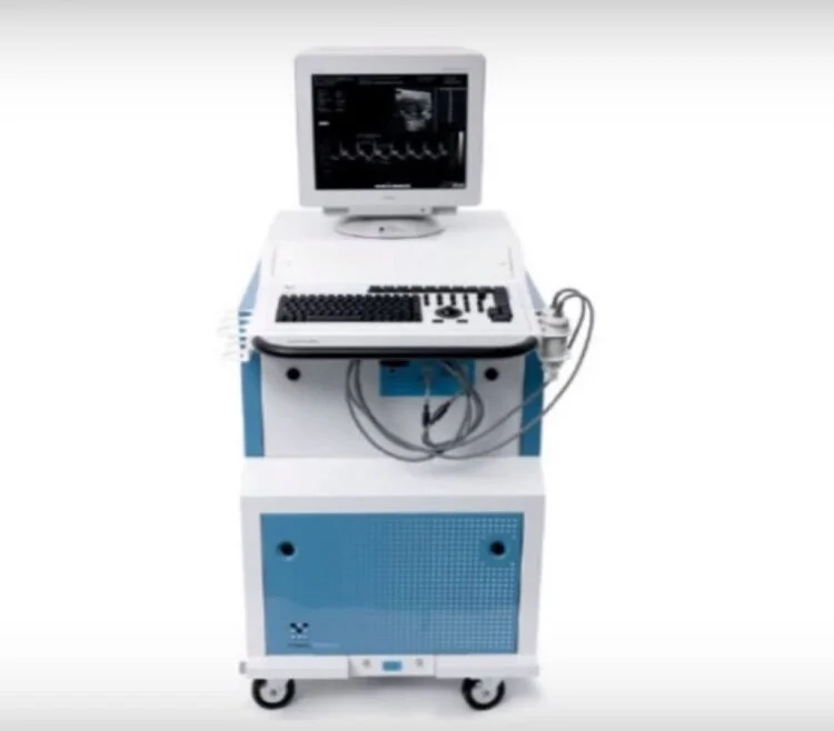
VisualSonics Vevo 2100 Micro Ultrasound
The Vevo 2100 Micro Ultrasound, in its most rudimentary application, can be used to produce a description of the anatomical structure of the cerebrovasculature to a depth of 10 mm from the brain surface and spatial resolution down to 15 µm (when used in conjunction with microbubbles). Investigation on perfusion is also possible as the frame rate can reach up to 1000 frames/s producing a nearly live feed of blood flow through the cerebrovasculature. Finally, compared to other imaging modalities available, micro-ultrasound is non-invasive, relatively cost-effective and portable. Find more info here.