FACILITY EQUIPMENT
MULTIPHOTON LASER SCANNING MICROSCOPE SYSTEMS

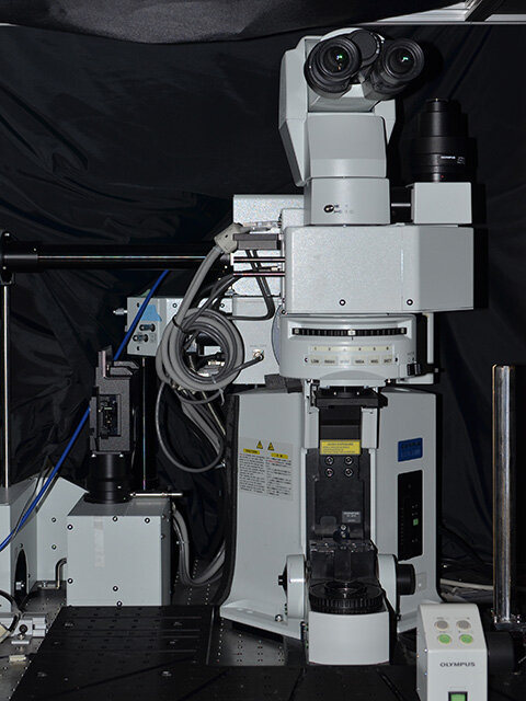
Olympus FVMPE-RS
The newer FVMPE-RS system allows high speed scanning (up to 30 frames per second for a 512x512 full field of view; or 438 frps at 512x32) with an optical path optimized for near-infrared transmission, allowing deeper imaging into the brain. Light is sourced from two Spectra Physics’ Titanium:sapphire lasers (MaiTai and Insight models for concurrent deep red imaging (680-1300nm) and photostimulation for optogenetic applications. The microscope is also equipped with visible light photostimulation capabilities (458 and 552nm), sequential scan management for complex imaging paradigms, and triggering systems for integration with other lab equipment. Find more technical information here.
Olympus FV1000MPE
The Olympus FV1000MPE upright multiphoton laser scanning microscopy system is equipped with an IR Mai Tai HP mode-locked Titanium Sapphire DeepSee tunable laser (690–1040 nm, Spectra Physics) for multiphoton imaging and with visible laser lines (405 nm, 440nm, 488nm, 514nm, 561n, and 635nm) for confocal imaging. Emitted flourescence is collected using four external photomultiplier tubes (Hamamatsu Photonics). The system is used to scan fixed or live images, and features include Z-stack 3D imaging and time series up to 16 frames/sec. Find full technical information here.
WIDEFIELD FLUORESCENCE MICROSCOPE

Zeiss Axiovert 200M Inverted Fluorescence Microscope
The Zeiss Axiovert 200M is an inverted fluorescence microscope with brightfield capability which is suitable for multi-channel fluorescence imaging of fixed samples, in addition to basic live-cell fluorescence imaging and transmitted light imaging.
Additional Imaging Equipment
We regularly utilize the Nikon A1 Laser Scanning Confocal Microscope and the Zeiss Spinning Disk Microscope from the SRI Centre for Flow Cytometry and Scanning Microscopy (CCSM) located in M-Wing.
ELECTROPHYSIOLOGY EQUIPMENT

Tucker-Davis Technologies 32-Channel System
Electrophysiology acquisition system for extracellular recordings (local field potential/single unit); 32 channel (2 x 16). Synchronous stimulation for electrical (4x4 channels) and light pulses, with switching headstage for electrical stimulation and recording on the same electrode by fast-switching (<0.2 ms) between the two modes.
Neuropixels 1.0 Probe
The Neuropixels 1.0 probe is a fully-integrated silicon CMOS digital neural probe with on-chip circuitry for signal conditioning and digitization. The probe features 384 dual-band, low-noise recording channels that can individually be configured to simultaneously record AP (action potential) and LFP (local field potential) signals from 960 selectable, low-impedance TiN electrodes densely tiled along a 10-mm long, 70 x 24 µm cross-section straight shank. Voltage signals are filtered, amplified, multiplexed and digitized on-chip, allowing the direct streaming of digital data from the probe.

HEKA EPC10 Double Patch Clamp Amplifier
Continuing the tradition of providing the world's best electrophysiology amplifiers, the EPC 10 USB Double patch clamp amplifier is the optimal instrument for performing double patch experiments. Although two (EPC 10 USB Double) amplifiers are combined in a single housing; each amplifier is completely independent with clearly defined operation and handling. HEKA's software stimulates the desired amplifier and selected channels are programmed without tedious connection of cables by the user.
PRECLINICAL HIGH FREQUENCY MICRO ULTRASOUND IMAGING SYSTEM
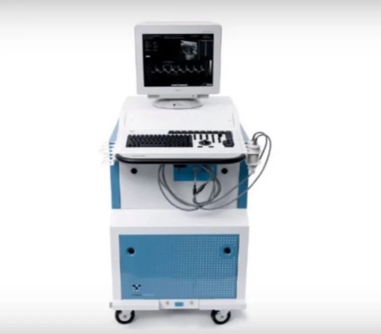
VisualSonics Vevo 2100 Micro Ultrasound
The Vevo 2100 Micro Ultrasound, in its most rudimentary application, can be used to produce a description of the anatomical structure of the cerebrovasculature to a depth of 10 mm from the brain surface and spatial resolution down to 15 µm (when used in conjunction with microbubbles). Investigation on perfusion is also possible as the frame rate can reach up to 1000 frames/s producing a nearly live feed of blood flow through the cerebrovasculature. Finally, compared to other imaging modalities available, micro-ultrasound is non-invasive, relatively cost-effective and portable. Find more info here.